
Ear feels blocked, hearing seems reduced, or your child is not responding to sounds clearly?


Middle ear fluid is one of the most common causes of hearing difficulty in children and is more frequently seen in adults than most people realise. The ear feels full, sounds seem muffled, and there is often no pain or fever to explain why.
Dr. Archana Jhawar, MS ENT with 25 years of experience, evaluates and treats middle ear fluid at Neoalta Clinic, Sector 17 Vashi and Kokilaben Hospital, Koparkhairane, Navi Mumbai.
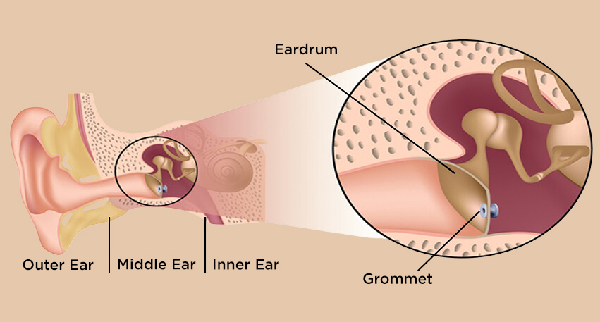
Middle ear fluid, medically called otitis media with effusion, is the collection of non-infected fluid in the middle ear space (normally air-filled cavity directly behind your eardrum).
The middle ear is connected to the back of the nose through a narrow channel called the Eustachian tube. When this tube does not work correctly, the air inside the middle ear gets absorbed and is replaced by fluid produced by the ear’s own lining.
This fluid is not infected, there is no pus, no bacteria, and no active illness. It is sticky and thick, which is why this condition is commonly called glue ear.
Its presence dampens the movement of the eardrum and the three small bones that conduct sound, causing the muffled or blocked sensation that is the main complaint in both children and adults.
Middle ear fluid can affect one ear or both and may remain for weeks to months before clearing on its own. In children it is the most common cause of mild to moderate temporary hearing difficulty in the early years of life.
This is the most common source of confusion for patients and parents. Both conditions involve the middle ear and both cause a blocked sensation and reduced hearing, but they are quite different conditions with different causes, symptoms, and treatment.
Ear infection involves active bacterial or viral infection inside the middle ear.
There is pus or infected fluid, significant pain, fever, and sometimes spontaneous discharge from the ear if the eardrum bursts under pressure. It comes on quickly, usually during or just after a cold, and is clearly uncomfortable.
Glue ear involves non-infected, sticky fluid. There is no active infection, typically no pain, and no fever.
The main complaint is a feeling of fullness or blockage in the ear with muffled hearing. In children it is often not noticed as a hearing problem, it shows up as inattentiveness, louder television use, or difficulty following instructions.
A key point: a child or adult who has had repeated ear infections may develop middle ear fluid as the infections clear. The infection resolves, but fluid lingers behind the eardrum for weeks or months afterward.
You can also read about Ear Infection Treatment in vashi.
read more about Ear infection-symptoms and treatment in this blog
The Eustachian tube is responsible for draining fluid from the middle ear and maintaining equal air pressure on both sides of the eardrum. When this tube is blocked or fails to open properly, fluid accumulates in the middle ear space.
Children are far more prone to middle ear fluid than adults, primarily because of differences in anatomy and immunity:
Middle ear fluid in adults is usually triggered by one of the following:
Yes, middle ear fluid is not just a childhood condition. Adults can also develop it.
Adults with persistent ear blockage often assume wax is the cause, but fluid behind the eardrum is another common reason for reduced hearing after a cold, allergy flare-up, or flight.
The most common description from adult patients is: ‘My ear blocked during a cold and never fully unblocked’ or ‘My ears did not pop after the flight and it has been three weeks.’
In most adults, middle ear fluid following a cold or flight resolves naturally within 4–6 weeks as the Eustachian tube recovers and the fluid drains on its own. No specific treatment is needed during this period beyond treating any underlying nasal congestion or allergy.
If the blocked sensation and reduced hearing persist beyond 6–8 weeks in an adult without clear improvement, ENT evaluation is recommended.
Tympanometry will confirm whether fluid is still present.
In adults, particularly when only one ear is affected and there is no obvious preceding cause, a visit to an ENT specialist and a thorough nasal and nasopharyngeal examination is important.
Diagnosis of fluid in the middle ear is accurate and straightforward when the right tests are performed together.
The eardrum is examined with a camera that displays a real-time image on a monitor so you can see exactly what is happening inside your own ear.
In middle ear fluid, the eardrum appears dull and slightly yellowish or amber rather than its normal pearly-bluish colour. Air bubbles or a visible fluid level behind the eardrum may sometimes be seen directly.
Tympanometry is a simple, painless in-clinic test that takes only a few minutes and requires no active participation from the patient.
A small probe is placed gently in the ear canal and air pressure is varied. The test measures how well the eardrum moves in response to pressure changes.
A flat line result called a Type B tympanogram, confirms the presence of fluid in the middle ear. It is the single most reliable test for diagnosing middle ear fluid and is available at Neoalta Clinic, Vashi.
A formal hearing test to measure how much the fluid is affecting hearing at different frequencies. It documents the degree of hearing loss, helps guide treatment decisions, and provides a baseline to compare against after treatment.
Since most cases of middle ear fluid are related to nasal or Eustachian tube dysfunction, ENT specialist examine the nose routinel. This may include nasal endoscopy to directly assess the opening of the Eustachian tube, size of the adenoids, nasal polyps, or septal deviation all of which can contribute to tube blockage.
In the majority of cases, yes. Most episodes of middle ear fluid, especially those following a cold or respiratory infection, resolve spontaneously within a few weeks.
During this period, regular follow-up, hearing assessment when indicated, and treatment of contributing factors such as allergy, nasal congestion, sinus disease, or Eustachian tube dysfunction may be recommended.
If fluid persists and is associated with hearing loss, speech concerns, or significant symptoms, further treatment options are discussed.
Treatment is chosen based on the duration of fluid, the degree of hearing loss, the patient’s age, and whether an underlying cause has been identified.
At Neoalta Clinic, Vashi and Kokilaben Hospital, Koparkhairane, all treatment decisions are made after complete ENT assessment by Dr Archana Jhawar.
First step is to diagnose and treat the underlying cause such as nasal allergy, sinusitis, chronic nasal congestion, or Eustachian tube dysfunction.
Nasal corticosteroid sprays, allergy medication, saline nasal irrigation, or sinus treatment may be recommended depending on what is Causing the fluid accumulation.
Treating the nose and Eustachian tube is often the most important step and in many patients, fluid clears once the root cause is addressed.
At our allergy clinic in Vashi, patients with recurrent middle ear fluid driven by allergy can be evaluated with skin prick testing and offered long-term treatment including SLIT (sublingual immunotherapy drops) or SCIT (allergy injections) when appropriate.
Regular follow-up allows the treating ENT to track whether fluid is clearing, hearing is improving, and whether further intervention is needed.
In selected patients, autoinflation technique (Valsalva)- gently blowing through the nose while pinching it closed may support Eustachian tube opening.
When fluid persists and is associated with documented hearing loss, significant symptoms, speech concerns, or impact on daily functioning, a myringotomy with ventilation tube (grommet insertion) may be advised.
A grommet is a tiny ventilation tube, inserted through the eardrum. It bypasses the blocked Eustachian tube, allows air into the middle ear, and lets the fluid drain out.
Hearing improvement is typically immediate after tube placement.
In children with recurrent or persistent middle ear fluid, enlarged adenoids are a common contributing cause.
Adenoid removal (adenoidectomy) at the time of grommet insertion significantly reduces the rate of recurrent fluid and the need for repeat grommet procedures.
Dr. Archana Jhawar performs adenoid surgery using coblation and microdebrider techniques at Kokilaben Hospital, Navi Mumbai. These techniques allow precise removal of adenoid tissue while minimizing injury to surrounding structures.
Middle ear fluid is not a medical emergency, but certain situations require prompt ENT assessment:
Middle ear fluid is one of the most common reasons children are brought to a paediatric ENT specialist. As there is no pain, fever or the clear distress of an ear infection, it can go unnoticed for weeks or months.
What parents most commonly notice first is not a hearing complaint from the child but a change in behaviour.
The child seems inattentive, keeps the television louder, does not respond when called from another room, or has difficulty following instructions in a noisy environment.
Teachers sometimes raise concerns about attention or listening before parents connect the behaviour to the child’s ears.
For most children, middle ear fluid is a temporary condition that clears naturally within a few weeks to months.
The critical period of concern is prolonged fluid beyond 3 months, in children under 5 years of age where the impact on speech and language development is most significant.
The decision to recommend grommet surgery is made carefully and is based on several factors together:
It is important for parents to understand that grommets do not cure the underlying Eustachian tube problem, they provide a temporary bypass while the child’s anatomy matures and immunity develops. Underlying allergy or enlarged adenoids are to be treated simulteneouly.
Serving patients from Vashi, Nerul, Belapur, Kharghar, Koparkhairane, Sanpada, Panvel, Airoli, Ghansoli and across Navi Mumbai.